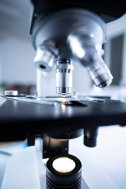
A microscope is a vital tool in microscopy, enabling detailed observation of tiny specimens. Understanding its parts and functions is essential for proper usage and optimal results.
1.1 Importance of Understanding Microscope Components
Understanding microscope components is crucial for effective use and maintenance. Each part plays a specific role in image formation and clarity. Knowing their functions ensures proper handling, preventing damage and optimizing performance. Misuse can lead to poor image quality or equipment harm. Grasping the basics enhances observational accuracy, especially in scientific and educational settings. Proper knowledge also aids in troubleshooting issues like focus problems or inadequate illumination. Familiarity with components like eyepieces, objectives, and condensers ensures efficient specimen analysis. This foundational understanding is vital for both beginners and experienced users, fostering confidence and proficiency in microscopy applications across biology, medicine, and research. Regular practice and review further solidify this essential skill.
1.2 Brief Overview of Microscope Types
Microscopes come in various types, each designed for specific applications. The compound microscope is the most common, using a combination of lenses for high magnification. Stereo microscopes provide 3D images, ideal for examining larger specimens. Electron microscopes use electron beams to achieve ultra-high resolution, essential for detailed cellular structures. Fluorescence microscopes highlight specific structures within specimens using fluorescent dyes. Darkfield microscopes enhance contrast for unstained samples. Each type caters to different needs, from routine laboratory work to advanced research. Understanding the strengths of each microscope type allows for appropriate selection based on the specimen and desired outcomes. This variety ensures microscopy remains versatile and effective across diverse scientific fields and applications.
Major Components of a Compound Microscope
The compound microscope consists of key parts: eyepiece, objective lenses, stage, illumination system, condenser, and focus knobs. These components work together to magnify and clarify specimens effectively.
2.1 Eyepiece (Ocular Lens)
The eyepiece, or ocular lens, is located at the top of the microscope. It magnifies the image produced by the objective lens, typically offering a 10X magnification. In binocular microscopes, two eyepieces are present, one for each eye, ensuring a clear and comfortable viewing experience. The eyepiece works by receiving the light from the objective lens and further enlarging the specimen’s image, making it visible to the user. Proper adjustment of the eyepiece, such as focusing, is crucial for obtaining a sharp and detailed view. Regular cleaning and maintenance of the eyepiece are recommended to prevent dust or debris from interfering with observation. This component is essential for the overall functionality of the microscope, enhancing the user’s ability to study specimens with precision.
2.2 Objective Lenses
The objective lenses are crucial components of a microscope, mounted on a rotating nosepiece. They collect light from the specimen and form an image that the eyepiece further magnifies. Available in various magnifications (e.g., 4X, 10X, 40X, 100X), they allow users to observe specimens at different scales. The objective lens with the lowest power (e.g., 4X) is typically used first to locate the specimen, while higher powers provide greater detail. Proper alignment and focus are essential for clear images. Objective lenses must be handled with care to avoid damage, and their quality significantly impacts the microscope’s performance. They are indispensable for achieving accurate and detailed observations in microscopy, making them a fundamental part of the microscope’s functionality.
2.3 Stage and Stage Clips
The stage is a flat platform on the microscope where the specimen slide is placed for observation; It is typically equipped with stage clips to securely hold the slide in position, ensuring stability during viewing. The stage is often adjustable, allowing for precise movement of the specimen under the objective lens. Some microscopes feature a mechanical stage with knobs to move the slide horizontally and vertically, making it easier to focus on different areas of the specimen. The stage clips are spring-loaded or screw-adjustable, providing a firm grip on the slide without damaging it. Proper use of the stage and clips is essential for maintaining the integrity of the specimen and achieving clear, focused images under the microscope.
2.4 Illumination System
The illumination system is a critical component of a microscope, providing light to view specimens. It typically includes a light source, such as an LED or halogen bulb, located beneath the stage. The system ensures even light distribution through the condenser and onto the specimen. Adjusting the light intensity is essential for optimizing visibility, often achieved using a dimmer switch or diaphragm. Proper alignment of the illumination system is necessary to avoid shadows and ensure a clear, well-lit image. Modern microscopes often feature energy-efficient LED lights for brighter, longer-lasting performance. The illumination system is vital for enhancing the clarity and detail of microscopic observations, making it indispensable in scientific and educational settings.
2.5 Condenser
The condenser is a crucial part of a microscope’s illumination system, focusing light onto the specimen. Positioned beneath the stage, it collects and directs light from the source, ensuring even distribution. The condenser adjusts light intensity and focus, optimizing image clarity. It typically includes a diaphragm to regulate light entry and a focusing mechanism for precise alignment. Proper condenser adjustment is essential for achieving sharp, high-contrast images. In darkfield microscopy, the condenser scatters light to create contrast. Regular maintenance, like cleaning, ensures optimal performance. The condenser plays a key role in enhancing image quality, making it a fundamental component in microscopy for both educational and professional use.
2.6 Focus Knobs (Coarse and Fine Adjustment)
The focus knobs are essential for adjusting the microscope’s focus. The coarse adjustment knob moves the objective lens farther from or closer to the specimen, bringing it into the field of view. The fine adjustment knob makes precise focal adjustments, ensuring a sharp image. Together, they enable clear visualization of the specimen at varying magnifications. Proper use of these knobs is critical for achieving optimal focus and resolving fine details. Regular maintenance ensures smooth operation, while correct handling prevents damage to the microscope. These knobs are indispensable tools for both students and professionals in microscopy, aiding in accurate specimen observation and analysis.
Additional Parts of a Microscope
Additional components include the diaphragm, stage clips, and light source, which enhance functionality by controlling light, securing specimens, and providing illumination for clear observations;
3.1 Body Tube
The body tube connects the eyepiece and objective lenses, housing the optical system. It ensures proper alignment and light transmission, critical for clear specimen visualization and magnification. The length and design of the body tube are standardized to maintain focus and prevent distortion. Proper care is essential to avoid damage, as it directly impacts image quality. Regular cleaning and handling with care prevent scratches or misalignment, ensuring optimal microscope performance. The body tube’s structural integrity is vital for accurate observations in various scientific and educational settings. Its durability and precision engineering make it a cornerstone of microscope functionality. Understanding its role enhances overall microscopy skills and outcomes.
3.2 Arm and Base

The arm and base are structural components that provide stability and support for the microscope. The arm connects the microscope’s body to the base, allowing for secure handling and transportation. The base is typically heavy and sturdy, ensuring the microscope remains steady during use. Proper handling involves gripping the arm firmly while carrying the microscope, preventing accidental damage. The base also houses the illumination system and other essential controls, making it a critical foundation for the microscope’s functionality. The design of the arm and base prioritizes durability and balance, ensuring smooth operation and minimizing vibrations that could disrupt observations. These components are fundamental to maintaining the integrity and performance of the microscope in various laboratory and educational settings.
3.3 Revolving Nosepiece
The revolving nosepiece is a crucial component that holds multiple objective lenses, allowing seamless switching between different magnifications. Positioned below the stage, it rotates to align the selected objective lens with the specimen. This mechanism ensures quick and efficient changes in magnification without disturbing the specimen’s position. The nosepiece typically features several openings, each securely holding an objective lens. Its design enables smooth transitions between objectives, maintaining focus and reducing preparation time. Proper alignment and cleanliness of the revolving nosepiece are essential for optimal performance. It is a key feature that enhances the versatility and functionality of the microscope, catering to various observational needs in scientific and educational settings.
3.4 Eyepiece Tube
The eyepiece tube is a structural component that houses the eyepiece lens, securely holding it in place. It connects the eyepiece to the objective lenses, ensuring proper alignment for image formation. The tube is typically made of durable materials to maintain stability and prevent misalignment. Its design allows for smooth rotation and adjustment, enabling users to position the eyepiece comfortably. The eyepiece tube plays a crucial role in maintaining the optical pathway, ensuring light from the objective lens reaches the eyepiece accurately. Proper care, such as regular cleaning, is essential to prevent dust or debris from interfering with the image quality. This component is vital for the microscope’s functionality and user experience.
3.5 Diaphragm
The diaphragm is a crucial component located below the stage of the microscope. Its primary function is to regulate the amount of light that passes through the specimen. By adjusting the diaphragm, users can control the aperture, allowing more or less light to enter the microscope. This adjustment helps optimize the illumination for clear observations. The diaphragm typically consists of a rotating disk with different-sized holes, enabling precise control over light intensity. Proper use of the diaphragm ensures that the specimen is evenly illuminated, enhancing image quality and contrast. It is an essential part of the microscope’s illumination system, aiding in achieving the best optical performance for detailed observations.
Functions of Each Microscope Part
Each microscope part plays a specific role in enhancing observation, from magnification and illumination to focus and specimen handling, ensuring precise and detailed visualization of microscopic structures.
4.1 Magnification Process
The magnification process in a microscope involves the combined effort of the eyepiece and objective lenses. The eyepiece, typically providing 10X magnification, works in tandem with the objective lens, which can range from 4X to 100X. When light passes through the specimen, the objective lens creates an enlarged image, which is then further magnified by the eyepiece. This dual-lens system produces a compounded magnification effect, such as 400X when a 10X eyepiece is paired with a 40X objective lens. To achieve higher magnification, the user switches to a higher-power objective lens using the revolving nosepiece. This process allows for detailed observation of microscopic structures, enhancing the visibility of tiny specimens for accurate analysis and study.
4.2 Light Transmission and Focus
Light transmission and focus are critical for achieving clear microscopic images. The illumination system provides light, which passes through the condenser and diaphragm to reach the specimen. The condenser focuses the light, ensuring even distribution, while the diaphragm adjusts light intensity for optimal contrast. The objective lens then collects and focuses this light to form an image. Focus knobs—coarse and fine adjustments—are used to align the specimen with the objective lens, ensuring sharpness and clarity. Proper adjustment of these components is essential for resolving fine details and obtaining accurate observations under the microscope.
4.3 Specimen Handling
Specimen handling is crucial for effective microscopic observation. The stage holds the specimen in place using stage clips, ensuring stability during viewing. Proper preparation, such as placing the specimen on a slide and covering it with a coverslip, prevents damage and contamination. The focus knobs adjust the lens height to bring the specimen into sharp focus. Handling slides gently and avoiding excessive pressure is essential to maintain the integrity of the specimen. Correct specimen placement and alignment under the objective lens are vital for accurate observations. This process ensures that the microscope functions optimally, providing clear and detailed images for analysis.
Understanding microscope components is essential for optimal performance and clear observations, making it a fundamental skill in scientific and educational microscopy applications.
5.1 Summary of Key Components
The microscope comprises essential parts that work together to enhance observation. The eyepiece magnifies the specimen, while objective lenses focus light. The stage holds the specimen securely, and the illumination system provides light. The condenser and diaphragm optimize light intensity and focus. Focus knobs adjust the image clarity, and the body tube houses the optical system. The arm and base provide stability, while the revolving nosepiece allows objective lens changes. The eyepiece tube ensures proper alignment, and the diaphragm regulates light entry. Each component plays a vital role in achieving precise magnification and clear visualization of specimens.
5.2 Importance of Proper Usage
Proper usage of a microscope ensures optimal performance, accuracy, and longevity of the instrument. Correct handling prevents damage to delicate components like lenses and the stage. Regular maintenance, such as cleaning and aligning parts, is crucial for clear observations. Misuse can lead to blurry images, inaccurate results, or even permanent damage to the microscope. Understanding and following the manufacturers’ guidelines helps in achieving precise focus and illumination. Proper techniques also enhance safety, avoiding eye strain or accidental breakage of slides. By adhering to best practices, users can maximize the microscope’s capabilities and extend its operational life, ensuring reliable outcomes in scientific and educational settings.